
IBBR Associate Director Co-Authors Science Paper on Vitamin A Transport
Thu, Sep 29, 2016
Using a new, lightning-fast camera paired with an electron microscope, University of Maryland School of Medicine scientists have captured images of one of the smallest human proteins to be “seen” with a microscope.
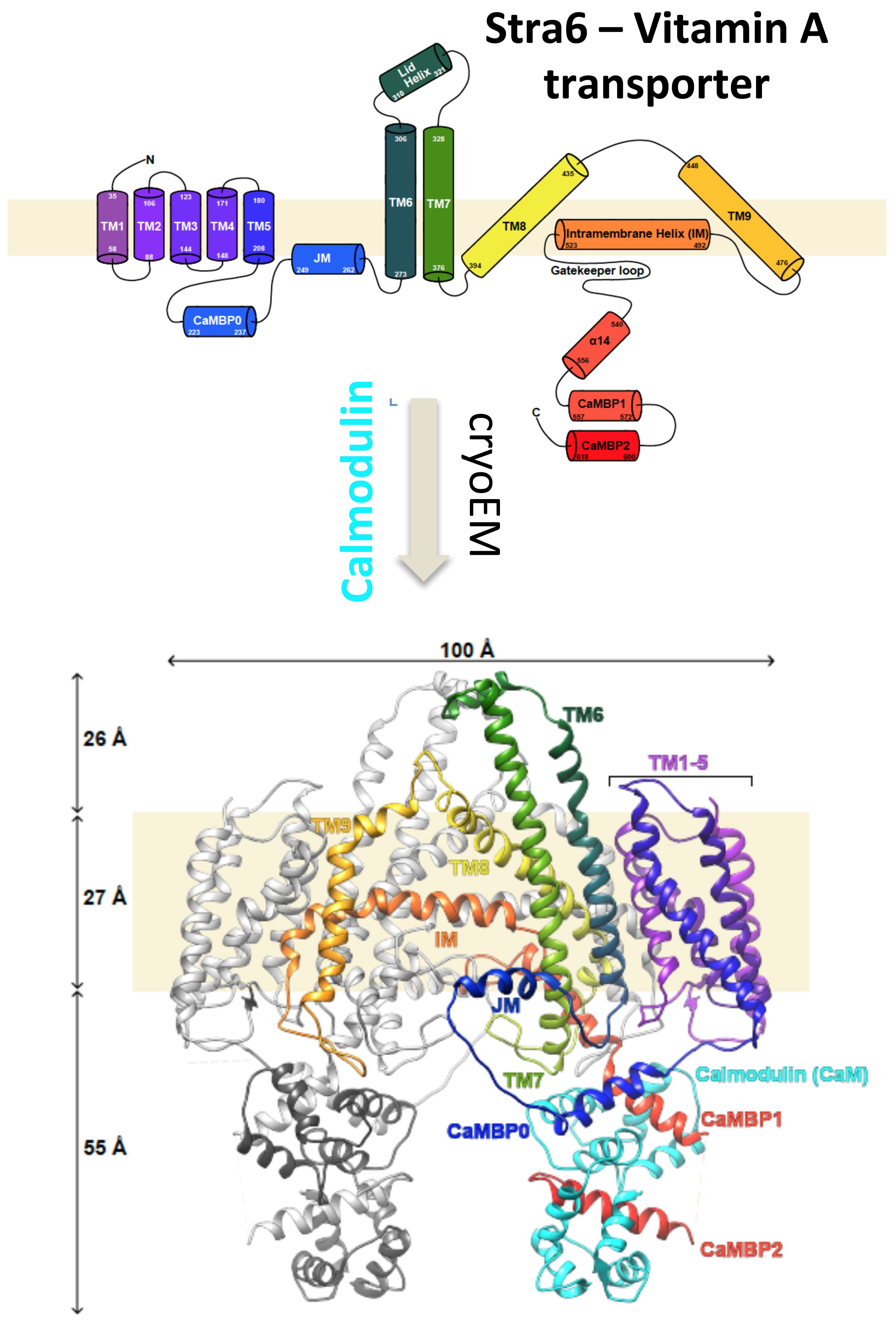
The protein – called STRA6 – sits in the membrane of our cells and is responsible for transporting vitamin A into the cell interior. Vitamin A is essential to all mammals and is particularly important in making the light receptors in our eyes, and in the placenta and fetus where it’s critical for normal development.
“Being able to visualize this protein, and understand how it works to move Vitamin A, is a really fantastic leap,” said one of the paper’s co-authors, David J. Weber, PhD, professor of biochemistry and molecular biology at the University of Maryland School of Medicine (UM SOM). “And there is so much more we can do with this technique. It’s exciting.”
Images of the protein, which revealed several unusual features, were published in the August 26 issue of the journal Science. The work is a collaboration among several scientists around the country. Structural biologist Filippo Mancia, PhD, assistant professor of physiology and cellular biophysics at Columbia University Medical Center, led a team of other scientists, including Dr. Weber of UM SOM, along with Wayne Hendrickson, Larry Shapiro, Joachim Frank and Bill Blaner at Columbia University Medical Center, Loredana Quadro at Rutgers University, and Chiara Manzini at George Washington University.
Until the new study, the way STRA6 transports vitamin A into the cell had been a mystery. Unlike most transporters, which interact directly with the substances they transport, STRA6 uses an intermediary protein that carries vitamin A in the bloodstream. Revealing the structure of STRA6 may provide insight into how other, related transporters work.
A new type of camera technology was a key element to getting the STRA6 images. When paired with an electron microscope, the camera allows biologists to see tiny, never-seen-before structural details of the inner machinery of our cells.
“We can now get near atomic resolution because the new camera is much faster and allows us to take a movie of the molecules,” says Oliver Clarke, PhD, an associate research scientist in the Hendrickson lab at Columbia University Medical Center. “Even under the electron microscope, the molecules are moving around by a tiny amount, but when you take a picture of something moving, it comes out blurry. With such a movie, we can align the frames of the movie to generate a sharper image.”
The researchers used approximately 70,000 individual pictures of STRA6 to generate a 3-dimensional map of the protein, which was used to construct an extremely accurate atomic model. The images and model reveal that STRA6 is “a bit of a freak,” says Dr. Clarke.
Though this needs to be verified, the mechanism may be a way to protect cells from too much vitamin A. “Vitamin A is actually somewhat toxic,” says Dr. Mancia. “Trapping vitamin A inside the membrane may keep control of the amount inside the cell.” The research may help researchers understand how other, still mysterious cellular components, work.
“This collaboration among research institutions has yielded fascinating insight into the cellular pathway of vitamin A,” said UM SOM Dean E. Albert Reece, MD, PhD, MBA, who is also vice president for medical affairs at the University of Maryland and the John Z. and Akiko K. Bowers Distinguished Professor. “When scientists work together like this, great accomplishments occur. The technique developed here will clearly reap future discoveries in other domains as well.”
About the University of Maryland School of Medicine
The University of Maryland School of Medicine was chartered in 1807 and is the first public medical school in the United States and continues today as an innovative leader in accelerating innovation and discovery in medicine. The School of Medicine is the founding school of the University of Maryland and is an integral part of the 11-campus University System of Maryland. Located on the University of Maryland’s Baltimore campus, the School of Medicine works closely with the University of Maryland Medical Center and Medical System to provide a research-intensive, academic and clinically based education. With 43 academic departments, centers and institutes and a faculty of more than 3,000 physicians and research scientists plus more than $400 million in extramural funding, the School is regarded as one of the leading biomedical research institutions in the U.S. with top-tier faculty and programs in brain science, cancer, surgery and transplantation, trauma and emergency medicine, vaccine development and human genomics, among other centers of excellence. The School is not only concerned with the health of the citizens of Maryland and the nation, but also has a global presence, with research and treatment facilities in more than 35 countries around the world. http://medschool.umaryland.edu/
Columbia University Medical Center provides international leadership in basic, preclinical, and clinical research; medical and health sciences education; and patient care. The medical center trains future leaders and includes the dedicated work of many physicians, scientists, public health professionals, dentists, and nurses at the College of Physicians and Surgeons, the Mailman School of Public Health, the College of Dental Medicine, the School of Nursing, the biomedical departments of the Graduate School of Arts and Sciences, and allied research centers and institutions. Columbia University Medical Center is home to the largest medical research enterprise in New York City and State and one of the largest faculty medical practices in the Northeast. For more information, visit cumc.columbia.edu or columbiadoctors.org.