Profile

Kristen Varney
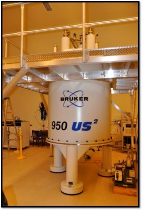
Dr. Varney has served as Manager of the University of Maryland, Baltimore Nuclear Magnetic Resonance (NMR) Shared Service Facility for over 14 years and as Structural Biology Section Leader for the Center for Biotherapeutics (CBT) since 2011. In addition to conducting her own research, as director of this facility, Dr. Varney promotes the use of NMR spectroscopy in University of Maryland School of Medicine NIH-funded research, manages all NMR-related projects conducted at the UMB NMR facility, and keeps the NMR spectrometers (600 MHz, 800 MHz, 950 MHz NMR) in good working condition.
A major component of Dr. Varney’s position is oversight of NIH-funded projects and training users. This training includes assisting investigators in preparing an appropriate NMR sample, troubleshooting sample conditions, setting up and running all NMR experiments, processing and analysis of NMR data, and use of all NMR-related computer software. In addition, she is skilled in implementing new NMR techniques for use in the facility, ensuring the best results for the >20 active projects currently in place at the facility.
CURRENT RESEARCH
Dr. Varney oversees numerous state-of-the-art scientific studies that utilize NMR spectroscopy to determine atomic resolution structures of proteins, protein complexes, and complexes involving biomolecular therapeutics and small molecule inhibitors. This work provides both structural and dynamic information useful in the investigation of mechanisms involved in disease states and the development of drugs to treat them.
Dr. Varney works collaboratively on multiple projects including an NMR binding study of KRas4b G12D (1-169) bound to compounds provided by Frank McCormick’s laboratory at University of California San Francisco. Mutated KRAS genes are drivers of colorectal cancer and exist in approximately 45% of colorectal cancer cases. Moreover, KRAS mutant colorectal cancers are intrinsically insensitive to epidermal growth factor receptor (EGFR) inhibitors. Anti-KRAS siRNA reduces proliferation and viability of colorectal cancer cell lines that are resistant to anti-EGFR therapy in vitro, and slows tumor growth in a xenograft model.
The current project involves the development of specific inhibitors of KRAS signaling that work via disruption of regulatory protein-protein interactions (PPIs) that drive the cascade. The development of specific KRAS inhibitors will enable the preclinical and clinical development of a novel class of cancer therapeutics and create the experimental infrastructure for additional targeted disruptions of protein-protein interactions that drive RAS signaling in cancer. Previously published NMR-based studies have successfully identified a KRAS-compound binding pocket where > 25 “hits” (including DCAI) interact with KRAS at the same location. Further, analysis of the crystal structure predicts that this binding pocket interferes with Ras/SOS interactions, thus potentially inhibiting SOS-mediated nucleotide exchange and preventing KRas activation (Maurer et al. 2012. PNAS).
The goal of this study is to use NMR chemical shift change analysis to identify novel KRAS inhibitors, estimate binding affinities, and determine whether the newly-identified inhibitors occupy the known KRAS binding site, or characterize a new binding site.
Publications
- Physiologically Relevant Free Ca2+ Ion Concentrations Regulate STRA6-Calmodulin Complex Formation via the BP2 Region of STRA6.
- 1HN, 13C, and 15N backbone resonance assignments of the SET/TAF-1β/I2PP2A oncoprotein (residues 23-225).
- The Importance of Therapeutically Targeting the Binary Toxin from Clostridioides difficile.
- Specificity of Molecular Fragments Binding to S100B versus S100A1 as Identified by NMR and Site Identification by Ligand Competitive Saturation (SILCS).
- 1HN, 13C, and 15N resonance assignments of the Clostridioides difficile receptor binding domain 2 (CDTb, residues 757-876).
- Correction to: 1HN, 13C, and 15N resonance assignments of human calmodulin bound to a peptide derived from the STRA6 vitamin A transporter (CaMBP2).
- Structure of the cell-binding component of the Clostridium difficile binary toxin reveals a di-heptamer macromolecular assembly.
- An asymmetry that leads to activity.
- Second harmonic generation detection of Ras conformational changes and discovery of a small molecule binder.
- 1HN, 13C, and 15N backbone resonance assignments of the human DNA ligase 3 DNA-binding domain (residues 257-477).
- 1HN, 13C, and 15N resonance assignments of human calmodulin bound to a peptide derived from the STRA6 vitamin A transporter (CaMBP2).
- Targeting S100 Calcium-Binding Proteins with Small Molecule Inhibitors.
- Identification of amino acid residues critical for the B cell growth-promoting activity of HIV-1 matrix protein p17 variants.
- Loss of S100A1 expression leads to Ca2+ release potentiation in mutant mice with disrupted CaM and S100A1 binding to CaMBD2 of RyR1.
- Kröhnke pyridines: Rapid and facile access to Mcl-1 inhibitors.
- A single amino acid substitution confers B-cell clonogenic activity to the HIV-1 matrix protein p17.
- The Activation of Protein Kinase A by the Calcium-Binding Protein S100A1 Is Independent of Cyclic AMP.
- Development of a glycoconjugate vaccine to prevent invasive Salmonella Typhimurium infections in sub-Saharan Africa.
- Crystal structure of the human heterogeneous ribonucleoprotein A18 RNA-recognition motif.
- Cellular aspartyl proteases promote the unconventional secretion of biologically active HIV-1 matrix protein p17.
- Structural basis of damage recognition by thymine DNA glycosylase: Key roles for N-terminal residues.
- (1)H(N), (13)C, and (15)N resonance assignments of the CDTb-interacting domain (CDTaBID) from the Clostridium difficile binary toxin catalytic component (CDTa, residues 1-221).
- 1H, 13C, and 15N resonance assignments of an enzymatically active domain from the catalytic component (CDTa, residues 216-420) of a binary toxin from Clostridium difficile.
- Small Molecule Inhibitors of Ca(2+)-S100B Reveal Two Protein Conformations.
- Chemical shift assignments for human apurinic/apyrimidinic endonuclease 1.
- Backbone assignment of HINT1 protein, a mouse histidine triad nucleotide binding protein.
- Small molecules bound to unique sites in the target protein binding cleft of calcium-bound S100B as characterized by nuclear magnetic resonance and X-ray crystallography.
- Solution structure of S100A1 bound to the CapZ peptide (TRTK12).
- S100A1 binds to the calmodulin-binding site of ryanodine receptor and modulates skeletal muscle excitation-contraction coupling.
- Restriction endonuclease inhibitor IPI* of bacteriophage T4: a novel structure for a dedicated target.
- Solution structure of the novel dispersin protein of enteroaggregative Escherichia coli.
- The three-dimensional solution structure of Ca(2+)-bound S100A1 as determined by NMR spectroscopy.
- Backbone dynamics of the olfactory marker protein as studied by 15N NMR relaxation measurements.
- Calcium-binding properties of wild-type and EF-hand mutants of S100B in the presence and absence of a peptide derived from the C-terminal negative regulatory domain of p53.
- Solution structure of zinc- and calcium-bound rat S100B as determined by nuclear magnetic resonance spectroscopy.